In this post
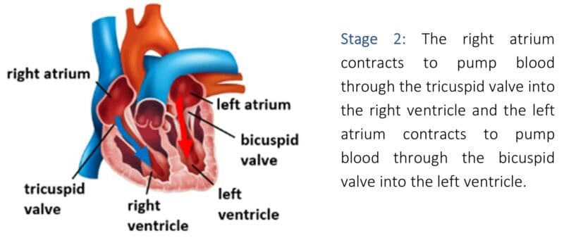
The human heart pumps blood around the body. The wall of the heart is made of cardiac muscle, enabling it to pump blood around the body at different speeds and pressures according to the body’s requirements. Cardiac muscle is unlike any other muscle in the human body as it does not tire like skeletal muscle. The following image shows an illustration of a labelled vertical section of the heart:

The four chambers of the heart are:
- The right atrium
- The right ventricle
- The left atrium
- The left ventricle
Cardiac muscle in the walls of the four chambers moves blood through the heart by a series of contractions and relaxations. The series of contractions and relaxations of the four chambers form what is known as the cardiac cycle. When a chamber of the heart is contracting, it is referred to as being in systole. When a chamber of the heart is relaxing, it is referred to as being in diastole.
Human heart valves
Before discussing the stages of the cardiac cycle, it is important that you recognise the main valves of the heart and their function. There are four valves in the human heart. These are:
- The pulmonary valve
- The tricuspid valve
- The aortic valve
- The mitral valve
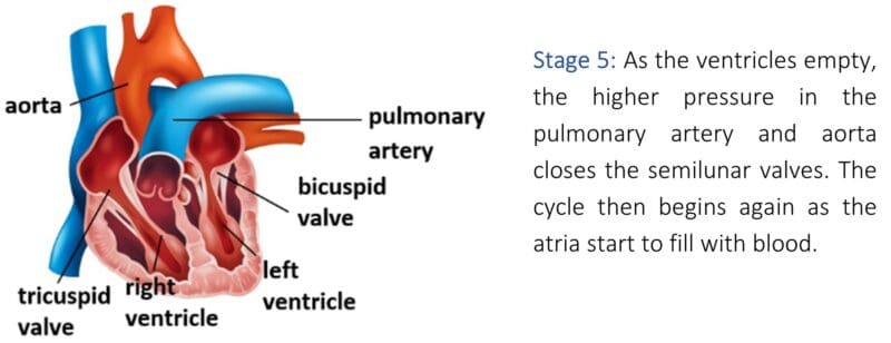
The diagram below illustrates these valves:

Heart valves ensure that the blood is flowing in the right direction. The pulmonary valve and the aortic valve are known collectively as semilunar valves. They are pocket-like structures that help prevent blood flowing backwards from the arteries to the ventricles during ventricular diastole (when the chambers in the heart are relaxing). The tricuspid valve separates the right atrium from the right ventricle and allows blood to flow into the ventricle but not backwards into the atrium. The mitral valve functions exactly the same as the tricuspid valve but on the left side of the heart. In other words, the semilunar valves control the blood flow coming out of the ventricles whereas the mitral valve and tricuspid valve control the blood flow from the atria to the ventricles.
As can be seen from the diagram, there are also such things as biological valves and mechanical valves. These valves are prosthetic models that are used to replace damaged and irreparable human heart valves.
Biological valves are replacement valves that are made from a biological source, for example from a human donor or from an animal (a pig heart is very similar to a human heart and so this is a common source of a biological valve if a human donor is not available). Mechanical valves are replacement valves that are man-made. Both types of artificial heart valves have different advantages and disadvantages:
| Advantages | Disadvantages | |
|---|---|---|
| Biological valves | These valves do not damage red blood cells as they pass through | These valves tend to last approximately 15 years and so for patients with a long-life expectancy, there is a high risk of further operations to replace the valves in the futureThese valves are prone to becoming hardened over several years |
| Mechanical valves | These valves are very strong and durable. Unlike biological valves, they are able to last a lifetime | These valves require the patient to take anti-blood clotting medication for the rest of their livesRed blood cells can become damaged as they pass through the valvesSome patients comment that they can hear the valves opening and closing |
Artificial hearts
There is also such a thing as an artificial (man-made) heart. An artificial heart provides an alternative to a human heart as it is able to replicate its function. It is used with patients that have severe heart disease, severe heart damage, or heart failure. An artificial heart is only often used when there is a shortage of a compatible human donor as current designs have not proven to be very successful in the long-term as they are prone to clotting within them. When there is a shortage of donors, many people die whilst they are on the waiting list, meaning that an artificial heart is often their only option until an appropriate donor becomes available.
The cardiac cycle
The main stages of the cardiac cycle are:


Coronary arteries
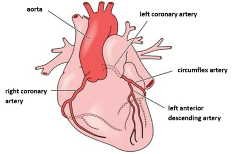
The coronary arteries provide the heart with its own blood supply. Two major coronary arteries branch off from the aorta before it meets the left ventricle (see left coronary artery and right coronary artery on the diagram below). These two arteries and their branches supply all parts of the heart muscle with blood.


