One of the organs that we use the most to detect stimuli is the eye. With sight we can locate objects, detect danger and ensure that we can make our way around. Although many species have eyes, very few have the same complexity as the human eye. For example, some animals may only be able to distinguish objects and only have eyes that can detect light, whereas others may be able to see objects but not colours. Here we will look at the complex organ of the eye and better understand how it uses light signals to build an image of our surroundings for the brain.

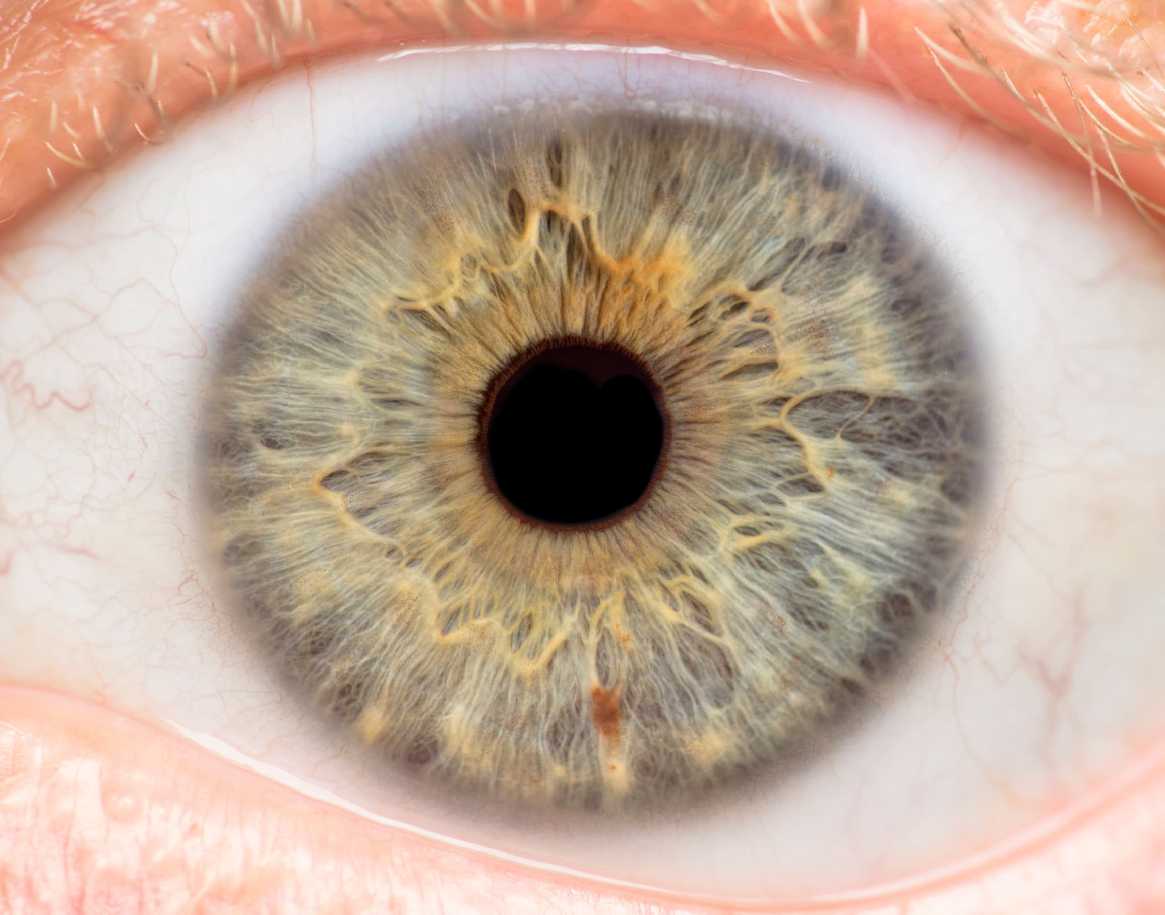
First off, the eye is protected by a tough outer layer known as the sclera. This is the visible, white section of the eyeball. Next there is the cornea which lets light into the eye to a thin layer of tissue called the iris. At the middle of the iris is the pupil which is a small hole that allows light through. The pupil is black because it does not give off any light and any light that goes into it is absorbed. All the way around the eye, within the sclera, is the choroid which is dark and contains lots of pigments and blood vessels. The reason for this is to stop light that has entered the pupil from being reflected around. The layer that is the furthest inside is the retina. This is the layer that is light sensitive and where the light energy that is taken into the eye is transformed into electrical impulses to be sent to the brain. The retina has special cells called rods and cones which react to light and produce impulses in sensory neurones that pass to the optic nerve. The rod cells work very well in poorly lit areas but cannot see different colours. So, when it is fairly dark, the rod cells work to allow us to see but not recognise colours. The cones only work in bright light and respond to different wavelengths of light which allows us to see in colour. These cones are concentrated at the back of the eye in a small depression known as the fovea. Cones give a much sharper image than rods do which is why our eyes work best when looking directly at an object – the light can fall into the fovea easily.
In order to form an image, the light that enters the eye must be refracted. Refraction is when light passes through one medium to another and is shifted when doing so. With the eye, the light that enters is refracted by the cornea and lens to flip the image on to the retina. The brain then takes this image and interprets it the right way up.

When you go from looking at something in dim lighting to something that is brightly lit, your eyes quickly adjust to this change. This is a good example of a reflex action. The purpose of this is to reduce the amount of light that falls on the retina as if this is too bright it could damage the cones and rods. However, if we don’t get enough light then we will struggle to form an image. Therefore, our eyes adjust automatically to allow the right amount of light through by changing the diameter of the pupil.
In the eye there is one area where we cannot form an image. This is known as a blind spot and is the area where the optic nerve leaves the eye. At this position there are no rods or cones and so the retina cannot see the image. However, as both eyes have a blind spot, the brain can cancel these out by using the image formed from one eye to compensate for the other.

