The human body’s cells get their energy through the process of respiration. In order to create the right amount of energy essential for life, cells are required to respire aerobically. For a cell to respire aerobically, oxygen must be present. The human body gets its oxygen through breathing. When we breathe, oxygen is drawn into the lungs. When oxygen is inhaled into the lungs, gas exchange between the oxygen and the blood can take place. Blood then carries this oxygen around the body to the cells and organs that require it. The cycle of ventilation is illustrated in the diagram below.
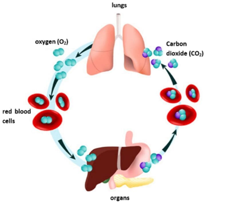
Breathing, also referred to as ventilation, is a different process to respiration. Respiration refers to the oxidation reaction occurring in cells. Be careful not to mix these terms up.
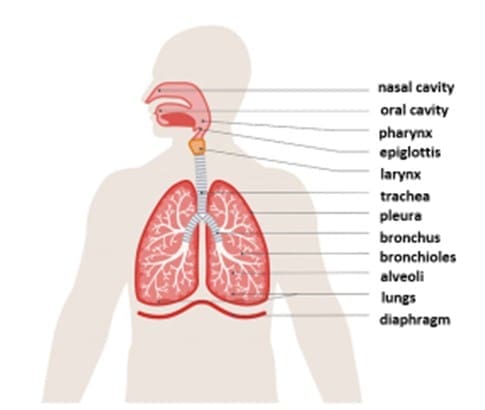
The nasal cavity is situated in the middle of the face and behind the nose. A nasal cavity is a large air space. There are two nasal cavities in the human body, each being a continuation of one of the nostrils. The oral cavity plays an important role in humans being able to digest food and in speech. However, we can also breathe through our mouths. When humans inhale (breathe in), air enters the nasal and/or the oral cavities and passes through another cavity known as the pharynx. The pharynx is a membrane-lined cavity that connects the nose and the mouth to the oesophagus. To ensure that we do not inhale food into our lungs, a piece of cartilage known as the epiglottis depresses during swallowing so that it covers the opening of the trachea (all structures from the neck to the diaphragm are found in what is called the human thorax). On the upper part of the trachea a muscular structure lined with a mucous membrane is located. This is known as the larynx and is where the vocal cords are. The trachea is a tube extending from the larynx to the bronchi (sing. bronchus) and is the principal passage of inhaling oxygen and exhaling CO2 from the lungs. Two thin, moist membranes known as pleura (or pleural membranes) surround each lung and separate them from the thorax. The pleural membranes envelope around each lung, making an airtight seal. Between the two membranes is a space known as the pleural cavity. This space is filled with a thin layer of pleural fluid. This liquid functions as a lubrication, preventing the surfaces of the lungs from sticking to the inside of the chest when we breathe.
The trachea branches into two bronchi (one bronchus in each lung). The bronchi divide into smaller tubes known as bronchioles which divide into smaller air sacs known as alveoli. The walls of the trachea and bronchi contain rings of cartilage. These rings of cartilage act to support the airways and keep them open as we breathe in.
The bronchial tree, as the name suggests, is a highly branching network of air passages. We breathe in from our nose or mouth, allowing air to pass down the trachea. The trachea splits into two tubes known as the bronchi; both lungs have one bronchus each. Both bronchi then divide into smaller tubes known as bronchioles which end in microscopic air sacs known as alveoli. Alveoli is where the gas exchange of oxygen and carbon dioxide with the blood takes place.
The diaphragm is the partition that separates the thoracic cavity from the abdominal cavity. The diaphragm has an important role to play in the process of breathing – we will discuss this in detail in the next chapter.
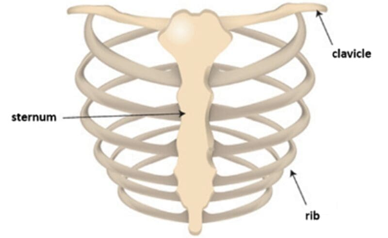
The thorax and the structures contained within the thorax are vital in a human’s ability to gain oxygen. Therefore, the inner organs contained within the thoracic cavity are protected by the clavicle, ribcage and the sternum. The clavicle, also known as the collar bone, is the only long bone in the human body that lies horizontally. There are two clavicles within the human body; one on the left and one on the right and they serve as struts between the sternum and the shoulder blades. The ribcage is a formation of slender curved bones known as ribs that surround the internal organs of the thoracic cavity. Rib bones are articulated in pairs to the spine; humans have 12 of them in total. The sternum, also known as the breastbone, is located in the centre of the chest forming the front of the ribcage. All of these bones help to protect the heart, lungs and the major blood vessels. Look at the following diagram which illustrates the bones protecting the thoracic cavity.
The role of intercostal muscles and the diaphragm in ventilation
Ventilation refers to the movement of air into and out of the lungs. Through the movement of breathing, we are able to change the volume of our thorax which allows for the difference in air pressure that is required to carry out the process of ventilation.
There are two movements that allow the thorax to change its volume which then leads to ventilation occurring. These movements involve the ribs and the diaphragm.
Intercostal muscles refer to the group of muscles that are situated between the ribs. Each rib is connected to the next by two sets of intercostal muscles. These muscles have an important function in helping to move the chest wall during inhalation and exhalation. The intercostal muscles are broken down into three layers. These are the external intercostal muscles, the internal intercostal muscles and the innermost intercostal muscles.
However, the diaphragm also works to move the chest wall during inhalation and exhalation. The diaphragm is situated at the bottom of the thoracic cavity and separates its contents from the abdominal cavity. It is a muscular, membranous wall that has a fibrous middle.
Both the intercostal muscles and the diaphragm play essential roles in the process of ventilation. The diagram below illustrates what happens during inhalation:
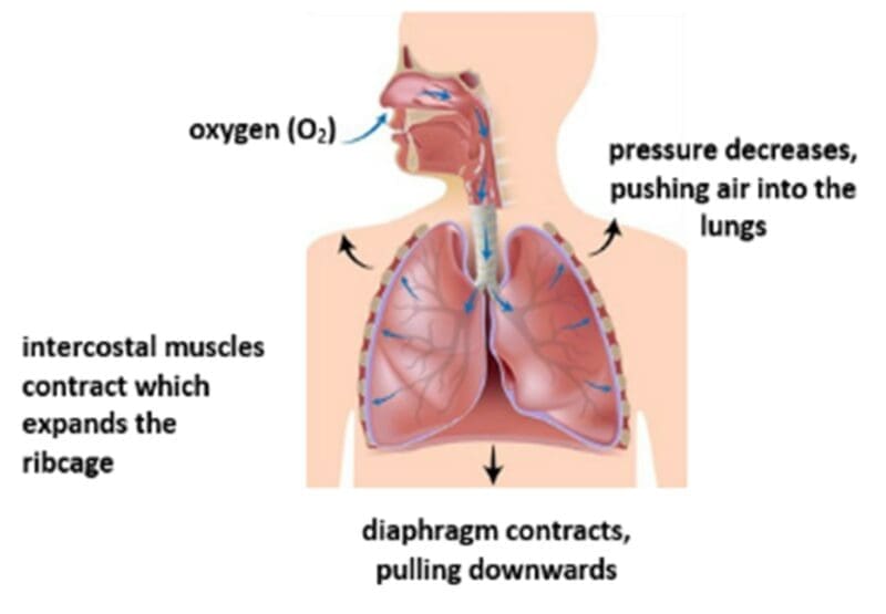
During inhalation, the ribcage is pulled upwards and outwards by the internal intercostal muscles relaxing and the external intercostal muscles contracting. At the same time, the diaphragm contracts, pulling it down. Air pressure inside the lungs decreases as lung volume increases and, as a result, air is pushed into the lungs. Most of the air pushed into the lungs is oxygen (O2), however, it will also contain some carbon dioxide (CO2) and some nitrogen (N).
The diagram below illustrates what happens during exhalation:
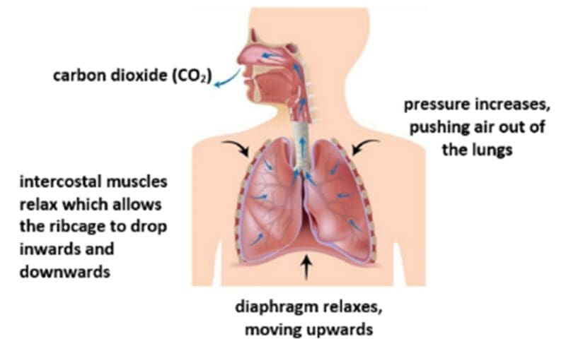
During exhalation, the ribcage is pulled downwards and inwards by the external intercostal muscles relaxing and the internal costal muscles contracting. At the same time, the diaphragm relaxes, moving upwards. Air pressure inside the lungs increases as lung volume decreases and air is pushed out of the lungs. Most of the air pushed out of the lungs is carbon dioxide (CO2), however, it will also contain some oxygen (O2) and some nitrogen (N). If you put your hands on your chest and breath in deeply, you will feel the movement of the ribs moving up and down.
Keeping the airways clean
It is important for the process of ventilation that the airways are kept clean and free from dirt particles and bacteria. A layer of cells lines the trachea and larger airways. These cells have a particularly important role in keeping the airways clean. In the lining, some of these cells secrete a thick and sticky liquid known as mucus. Mucus acts to trap particles of dirt or bacteria that are inhaled. Other cells in this lining are covered in tiny hair-like structure known as cilia. Cilia move backwards and forwards, acting like a sweeping brush to sweep the dirt particles, bacteria and mucus out towards the mouth. When dirt particles or bacteria enter the lungs then it can cause an infection, so mucus and cilia are particularly important in helping to prevent this.


