In this post
Large, multicellular animals require a circulatory system to transport substances to and from the cells in the body. The circulatory system, also known as the cardiovascular system, permits blood to circulate around the body so that it can provide cells with essential nutrients, help protect the body from disease and to also allow for the process of gas exchange.
The circulatory system is made up of the heart, blood vessels and blood. Blood is a complex tissue that flows through arteries and veins. As blood travels around the body, it collects materials from some places and deposits them in others. The substances that make up blood are:
- Plasma
- White blood cells
- Platelets
- Red blood cells
The following diagrams illustrate the composition of blood and their approximate measurements in reference to one another:


Plasma
Plasma constitutes approximately 52% to 62% of whole blood. Plasma is straw-coloured and is the main component of liquid in the blood. It is primarily responsible for transporting dissolved substances around the body. These include:
- Digested food (nutrients), for example water, glucose, amino acids, minerals and vitamins, from the gut to other parts of the body
- Waste substances, for example carbon dioxide, from all parts of the body to the lungs; and also urea from the liver to the kidneys
- Hormones
Plasma is also responsible for transporting heat energy around the body.
White blood cells
White blood cells, also known as leukocytes, constitute less than 1% of whole blood. They are an essential factor in the body’s immune system. There are several different types of white blood cells but their main roles are to engulf bacteria and other pathogens through the process of phagocytosis (literally meaning ‘cell eating’) and also to destroy harmful bacteria and pathogens through antibody production.
White blood cells can adapt and change shape easily. There are two main types of white blood cells: lymphocytes and phagocytes.

Lymphocytes make up approximately 25% of all white blood cells and their function is to produce antibodies. They have a large spherical nucleus and are approximately the same size as red blood cells. Lymphocytes produce antibodies to help destroy microorganisms. Pathogens, such as bacteria and viruses, have telltale chemical ‘markers’ on their surfaces known as antigens. Antibodies are able to recognise these antigens and stick to their surface to destroy the pathogen (known as a primary immune response).
Antibodies destroy pathogens in a number of ways. For example:
- They can cause bacterial cells to burst open
- They are able to neutralise poisons (toxins) produced by pathogens
- They can cause bacteria to stick together, making it easier for phagocytes to ingest them
- They act as a ‘label’ on the pathogen, making is more recognisable to a phagocyte
Antibodies – soluble proteins that help protect the body against pathogens, for example bacteria and viruses. Antibodies are produced by lymphocytes that are then passed into the plasma.
Some lymphocytes develop into memory cells. Memory cells are what make humans immune to certain diseases (known as the secondary immune response). When the same microorganism re-infects the body, a memory cell will start to reproduce and produce antibodies so the pathogen can be dealt with quickly. These cells can remain in the blood for many years and sometimes for someone’s entire life.
Phagocytes make up approximately 70% of all white blood cells. Phagocytes have a large spherical nucleus but sometimes this is a lobed nucleus. Phagocytes are also much larger than lymphocytes and red blood cells. They function to engulf bacteria and other microorganisms that have infected the human body through a process known as phagocytosis (known as a primary immune response):

Platelets
When the skin is broken or cut, there is a substantial risk that a person may lose a lot of blood or that harmful microorganisms could enter the bloodstream. Platelets, also known as thrombocytes, constitute less than 1% of whole blood. They are not whole cells and are instead fragments of large cells that are made in the bone marrow. The function of these cells is to stop bleeding when an injury occurs by helping the blood to clot and a scab to form.
When a break in the skin or a cut occurs, air stimulates the platelets in the blood and the damaged tissue to produce a chemical that causes fibrinogen (a soluble plasma protein) to form a network of insoluble fibres of fibrin (another type of protein) across the wound. This network traps platelets and red blood cells to form a clot, preventing pathogens entering the body and helping to prevent further blood loss. This clot then forms a scab which protects any damaged tissue whilst new skin grows underneath.
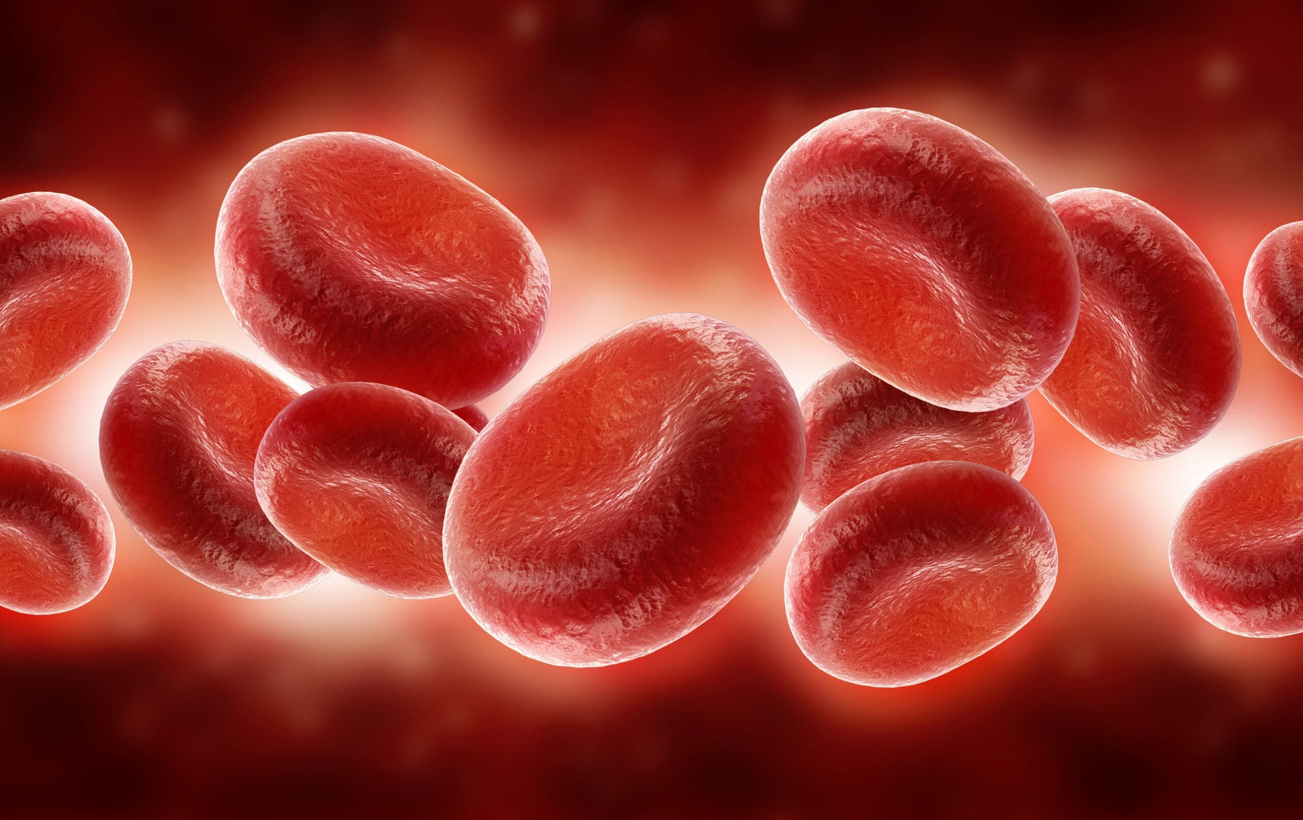
Red blood cells
Red blood cells, also known as erythrocytes, constitute approximately 38% to 48% of whole blood. They are highly specialised cells that are produced in the bone marrow. Their only function is to transport oxygen around the body. As red blood cells pass through the lungs, they load up with oxygen. As they pass through active tissues, they unload their oxygen.

The transport of oxygen is efficient because:
- Red blood cells are very small which allows them to pass through narrow capillaries
- There is a huge number of red blood cells (one drop of blood contains millions of red blood cells)
- Red blood cells contain a protein known as haemoglobin (an iron-containing protein). Haemoglobin absorbs oxygen in the lungs and carries it around in the blood before releasing it to the rest of the body. When there is a high concentration of oxygen surrounding haemoglobin it absorbs oxygen molecules to form a bright red oxyhaemoglobin. However, in oxygen-deficient environments, this process is reversed; in other words, when a low concentration of oxygen surrounds oxyhaemoglobin, it turns back into haemoglobin
- Red blood cells have a flattened disc shape to increase their surface area. Because of the thinness of the cell due to its flattened disc shape, it gives a short diffusion distance to the centre of the cell. They also have a large surface area to volume ratio, which allows for the rapid diffusion of oxygen when combined with their thin cell surface membranes
- Red blood cells do not have a nucleus. When red blood cells are being produced in the bone marrow, they lose their nucleus. This results in there being more space for haemoglobin which means more oxygen can be transported
Red blood cells have a limited life span of approximately 100 days; after this they are destroyed in the spleen.
Circulatory systems
Unicellular (single-celled) organisms, like the ones depicted below, do not have circulatory systems.

Because of their large surface area to volume ratio, unicellular organisms do not require a special system to transport materials around them. There is also no need for lungs or gills to obtain oxygen from the environment either as they can obtain their oxygen through their cell membrane; normally by the process of diffusion. The amount of oxygen that an organism can get (the supply rate) is determined by the area of the cell’s surface, and how much oxygen a cell uses is determined by the volume of the cell.
One of the primary functions of the circulatory system in animals is to transport oxygen. Blood is pumped to a gas exchange organ (either lungs or gills) to collect oxygen before it is pumped to the other areas of the body to unload this oxygen. There are two main types of circulatory system in animals:
- Single circulatory system – where the blood is pumped from the heart to the gas exchange organ and then straight to the rest of the body. Fish are an example of an animal organism that has a single circulatory system
- Double circulatory system – the blood is pumped from the heart to the gas exchange organ and back to the heart before being pumped to the rest of the body. Humans and other mammals are examples of animal organisms with double circulatory systems
The single circulatory system of a fish loses pressure as it passes through the gills, resulting in the oxygenated blood travelling slower to the rest of the organs. However, a double circulatory system in humans is much more efficient as it pumps blood twice. This results in higher pressures being maintained so the oxygenated blood can travel quicker to the organs and tissues of the body.
The human circulatory system consists of:
- The heart – the pump
- The blood vessels – these vessels carry blood around the body. Blood vessels refer to veins, arteries and capillaries. Veins carry the blood towards the heart; arteries carry the blood away from the heart; and capillaries carry the blood through the organs
- The blood – the transport medium
A double circulatory system consists of two distinct areas:
- Pulmonary circulation – where blood is circulated through the lungs where it is oxygenated
- Systemic circulation – where oxygenated blood is circulated to other tissues and organs of the body where it provides them with oxygen
The following diagram illustrates the double circulatory system in humans (the lines inside the red circles indicate the capillaries – we will discuss the structure and function of these in much more detail in a later chapter on this topic).


